
Figure 7. The degree of enteric innervation correlates with intestinal transit.
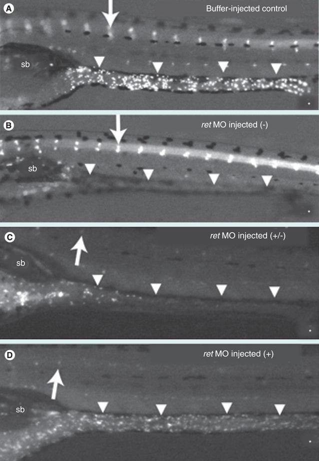
(A–D) Enteric neurons labeled with anti-HuC/D antibody are present in the cranial ganglia (arrow) in both buffer-injected control (A) and ret MO-injected (B–D) larvae. (A) In the buffer-injected control larvae, enteric neurons are present along the length of the GI tract (arrowheads). The majority of these larvae have completed or nearly completed transit after 24 h. (B–D) ret MO-injected larvae display variable degrees of enteric neuron loss along the length of the GI tract (arrowheads): no enteric neurons (−), reduced numbers of enteric neurons (+/−), wild-type enteric neurons (+). (C) Intestinal transit is delayed in ret MO-injected larvae with reduced enteric neurons. (D) ret MO-injected larvae with a normal complement of enteric innervation have a similar transit profile to the control larvae. The sb and anal opening (*) are indicated.
Figure obtained from Field et al. (2009) [100]. Reprinted from [100] with permission of John Wiley & Sons Inc.
MO: Morpholino; Sb: Swim bladder.