Figure 3.
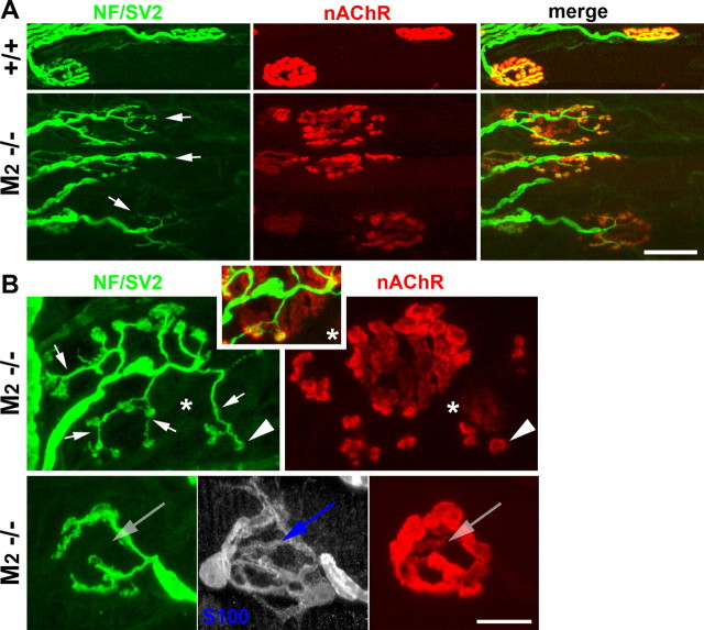
Spontaneous sprouting and terminal loss at M2−/− NMJs. A, Low-magnification confocal views of M2+/+ and M2−/− NMJs. Terminal arbors of many M2−/− NMJs (arrows; green) do not have the compact oval shape of wild-type NMJs but display unusually long and dispersed branches in contact with fragmented patches of nAChRs. B, High-magnification view of M2−/− NMJs. Terminal sprouts form varicosities directly apposed to islands of nAChR patches (arrowheads), whereas parental terminal branches that covered original synaptic contacts are retracted, leaving abandoned, faintly labeled branches of parental clusters of nAChRs (e.g., area marked by an asterisk). Some M2−/− NMJs with no sprouting exhibit vacant branches of nAChRs and tSCs that are unoccupied by terminal branches (arrows in bottom), indicating that terminal loss precedes terminal sprouting in M2−/− muscles. Scale bars: A, 30 μm; B, 10 μm. NF, Neurofilaments.

