
Figure 6.
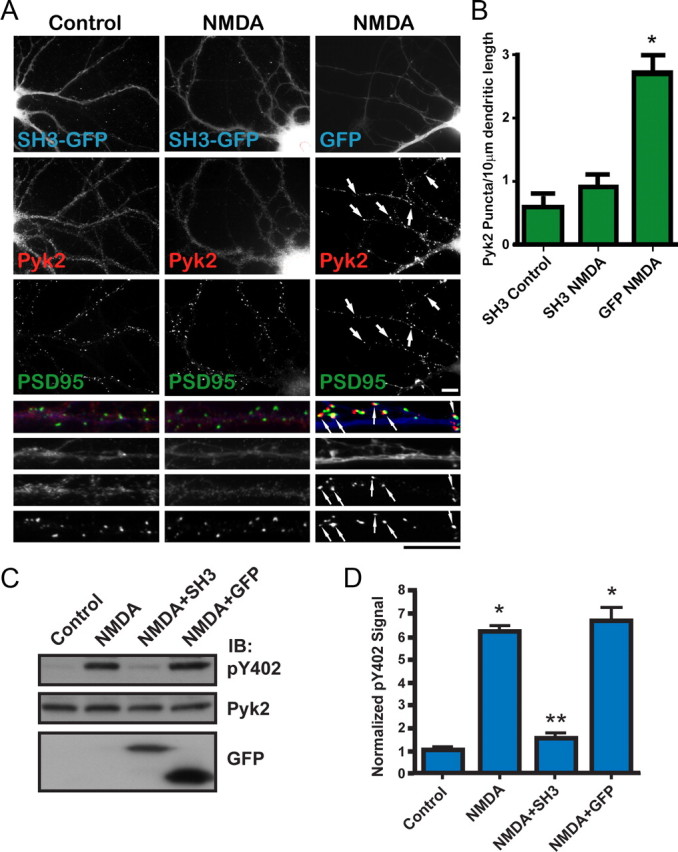
Overexpression of the PSD–95 SH3 domain inhibits NMDA-induced Pyk2 clustering and Pyk2 autophosphorylation. A, Primary hippocampal cultures (15 DIV) were transfected with the GFP-tagged SH3 domain of PSD-95 (SH3-GFP) or GFP alone (72 h; blue in overlay at higher magnification at bottom), treated with TTX plus vehicle or NMDA (50 μm, 5 min), fixed, and stained with antibodies against endogenous Pyk2 (αN; red in overlay) and PSD-95 (green in overlay). Scale bars, 10 μm for the corresponding magnifications. B, Density of Pyk2 immunoreactive puncta. SH3–GFP blocked NMDA-induced Pyk2 clustering seen in the presence of GFP alone. Five to 10 different fields were analyzed for each experiment, and values were averaged before statistical analysis for the means from the three independent experiments. The asterisk indicates statistical significance in Pyk2 clustering in neurons expressing GFP alone versus SH3–GFP (t test, p < 0.05; error bars indicate SEM). C, Primary hippocampal cultures (15 DIV) were infected with packaged FIV pVETL vectors encoding PSD-95 SH3–GFP or GFP alone (72 h) and treated with TTX plus pervanadate (1 mm) plus vehicle or NMDA (50 μm, 15 min) before immunoblotting (IB) with anti-phospho-Tyr402, monoclonal Pyk2, and GFP antibodies. D, Immunosignals were quantified by film densitometry. The pY402/Pyk2 ratios were normalization to control equaling 1. Asterisks denote statistical significance compared with control (*) or NMDA plus GFP (**) (t test, p < 0.05; n = 3 ± SEM).