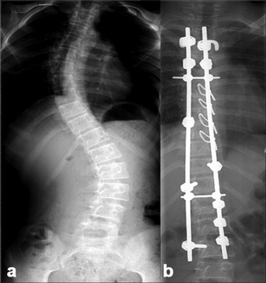
Figure 2.
(a) Preoperative X-ray of dorsolumbar and sacral spine (anteroposterior view) of a 12 year old female with Lenke Type1C curve. (b) Post-operative radiograph depicting hybrid constructs with screws at the bottom and hook-claws at the top and sublaminar wires at the concave apex, resulting in a good correction

