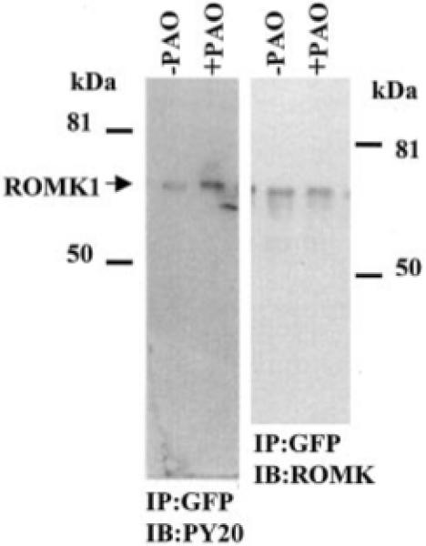
Fig. 5. A Western blot illustrating the effect of PAO on the tyrosine phosphorylation level of ROMK1 in the presence and absence of PAO.

The cells were treated with PAO or vehicle for 15 min. The ROMK1 channels were harvested by immunoprecipitation (IP) of the cell lysate with GFP antibody. The phosphorylated ROMK1 was detected with PY20 (left panel), and the total ROMK1 is recognized by ROMK antibody (right panel). The protein phosphorylation level is normalized by comparison to the relative amount of the total ROMK1 protein. IB, immunoblotting.
