
Figure 7.
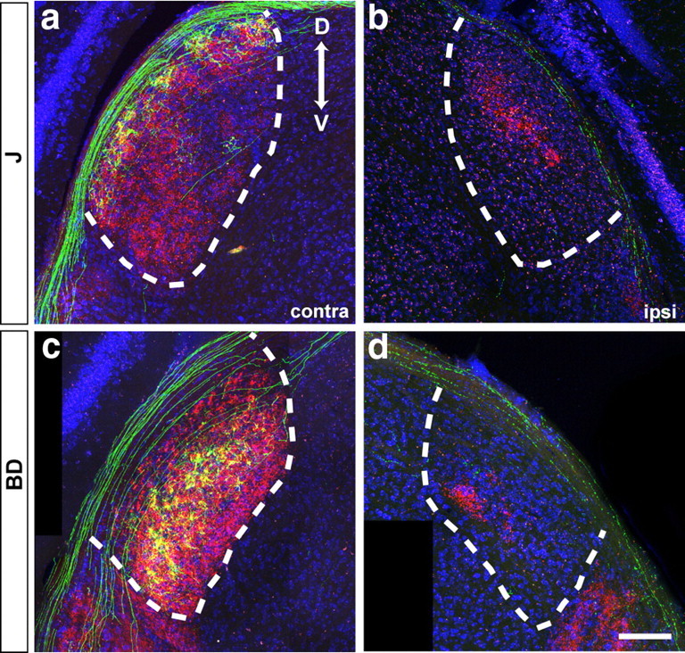
Lamina-restricted arbors of RGC axons in the lateral geniculate nucleus. Coronal sections were stained with anti-GFP (green) and Neurotrace (blue) after enucleation and cholera toxin administration (CTB, red). a, b, J-RGCs. c, d, BD-RGCs. Cholera toxin-positive regions in b and d contain projections from the ipsilateral retina. D, Dorsal, V: ventral. Scale bar: (in d) a–d, 150 μm.