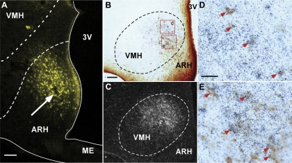
Fig. 6.
Afferent input into the ARH from CRFR2 neurons in the VMH. A: representative fluorescence micrograph showing fluorogold (FG) injection site (white arrow) in the ARH. B and C: representative photomicrographs showing FG-positive cells (B, dark brown color cells) and CRFR2 mRNA-positive signals (C, silver grain clusters) in the VMH. D and E: high-power magnification of boxed areas in B showing colocalization of FG immunoreactivity (brown color cells) and CRFR2 mRNA signals (black dot clusters). Representative double-labeled cells are indicated by red arrows. Scale bar = 125 (A), 100 (B), 25 (D) μm. ME, median eminence.

