Fig. 1.
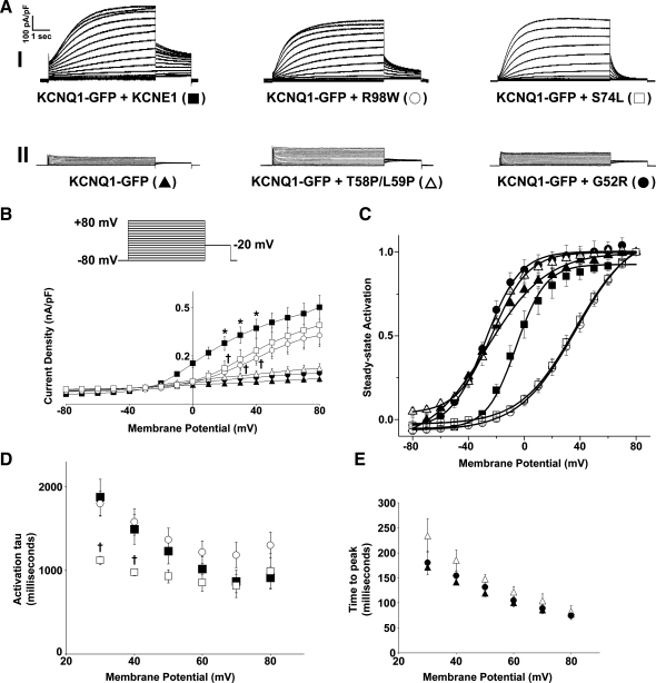
Differential effects of long QT syndrome type 5 (LQT5) mutants on the slow K+ current (IKs) channel function. A: representative traces of KCNQ1-green fluorescent protein (GFP) alone or in the presence of KCNE1 or the LQT5 mutants G52R, T58P/L59P, S74L, and R98W. Currents were normalized to cell capacitance, and the traces have been split into two categories: those that exhibit IKs characteristics (I) and those that do not possess the characteristics of IKs (II). The voltage protocol used is shown in the inset. B: mean current-voltage relationships. Current-voltage relationships were determined by normalizing the maximal current densities at the end of each pulse potential to cell capacitance (nA/pF). C: normalized voltage-dependent activation curves (steady-state activation) of KCNQ1-GFP expressed alone or in the presence of KCNE1 or the LQT5 mutants. The activation curves were determined by fitting the normalized amplitude of the peak tail currents versus test potential with a Boltzmann function (solid lines). D and E: mean activation time constants (activation τ) and time to peak values. These values were determined as described in materials and methods. Data are presented as means ± SE (n = 5–6 cells). *P < 0.05, significantly different from KCNQ1-GFP. †P < 0.05, significantly different from KCNQ1-GFP + KCNE1.

