
Figure 7.
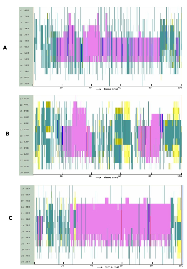
Evolution of secondary structures of the peptide variants at position 25 along the simulation: (A) p53: L25F (B) p63: F25L (C) p73: S25F; Colour code: purple, α-helix; red, π-helix; yellow, β-sheet; green, isolated bridge; cyan, turn; white, random coil.