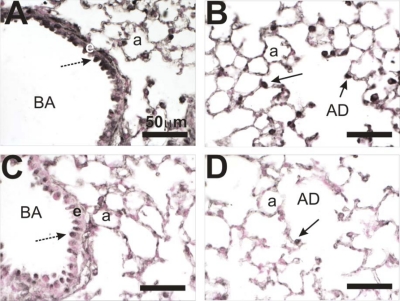
Fig. 1.
Hypoxia-inducible factor 1α (HIF1α) immunohistochemistry of lungs from control and doxycycline-treated mice. Lung tissue sections from control (A and B) and mice that were HIF1α deficient in their lungs (HIF1αΔ/Δ) (C and D) were analyzed by immunohistochemistry using a HIF1α-specific antibody. Control (A and B) and doxycycline (C and D)-treated mice were compared. HIF1α staining is prominent in the epithelial cell (e) lining the bronchiolar airway (BA) (dashed arrow) and type II cells (solid arrow) in the alveolar duct (AD) and alveolus (a). Staining is greatly reduced in the postnatally doxycycline-treated animals (C and D).

