Fig. 4.
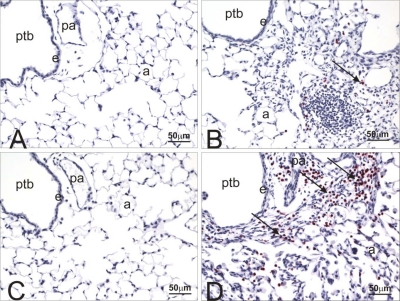
Major basic protein staining in lungs from control and HIF1αΔ/Δ mice. Light photomicrographs of the lungs of saline- (A and C) or cobalt-instilled (B and D) control (A and B) and HIF1αΔ/Δ (C and D) mice. All lung sections were immunohistochemically stained for major basic protein to identify infiltrating eosinophils (red chromagen; arrows) and counterstained with hematoxylin. A mixed inflammatory infiltrate consisting of mononuclear leukocytes, eosinophils, and lesser numbers of neutrophils are restricted to the lungs in cobalt-treated mice (B and D). Markedly more eosinophils are present in the peribronchiolar and alveolar regions of the cobalt-treated HIF1αΔ/Δ mouse (D) compared with that of the cobalt-treated control mouse (B). pa, Pulmonary arteriole; a, alveolar air space; e, bronchiolar epithelium.

