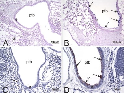
Fig. 5.
Alcian Blue/periodic acid-Schiff (AB/PAS) stain and YM1/2 immunohistochemistry (IHC). Light photomicrographs of ptb of cobalt-instilled control (A and C) and HIF1αΔ/Δ (B and D) mice. Tissues were stained with AB (pH 2.5)/PAS (A and B) to identify acidic and neutral mucosubstances (magenta stain; arrows) in mucous cells within the bronchiolar epithelium (e). Numerous AB/PAS-stained mucous cells are present only in the airway epithelium lining the preterminal bronchiole in the cobalt-treated HIF1αΔ/Δ mouse (B). Tissues in C and D were immunohistochemically stained for YM1/2 protein (brown chromagen; arrows) and counterstained with hematoxylin. YM1/2 proteins were present only in the bronchiolar epithelium of the cobalt-treated HIF1αΔ/Δ mouse (D). Arrow in C identifies a few alveolar macrophages that were positive for YM1/2 proteins.

