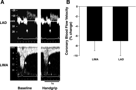
Fig. 1.
A: representative Doppler coronary blood flow velocity signal tracings from a patient during baseline (left) and static handgrip exercise (right) obtained by noninvasive transthoracic duplex ultrasound technique (TTD) from the left internal mammary artery (LIMA; bottom) grafted to the coronary artery and the left anterior descending artery (LAD; top). White arrows on LAD and LIMA tracings indicate the beginning of the diastolic component used to obtain mean coronary blood flow velocity. B: results are means ± SE, shown as percent change from baseline in coronary blood flow velocity during static handgrip at 50% of patient's respective maximum voluntary contraction. The measurements were obtained by noninvasive TTD simultaneously from the LIMA graft and the LAD. Note: the magnitude of decreases in coronary blood flow velocity was similar in 2 locations of the coronary artery.

