Figure 2.
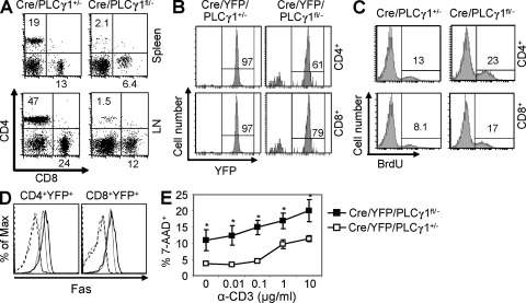
PLCγ1 deficiency results in T cell lymphopenia. (A) CD4/CD8 expression profiles on spleen and lymph node T cells from Cre/PLCγ1+/− and Cre/PLCγ1fl/− mice. Data represent six pairs of mice. (B) Percentages of YFP+ cells in splenic CD4+ and CD8+ populations from Cre/YFP/PLCγ1+/− and Cre/YFP/PLCγ1fl/− mice. Data represent five pairs of mice. (C) PLCγ1-deficient peripheral T cells displayed higher rates of BrdU incorporation. Data represent three pairs of mice. (D) PLCγ1-deficient T cells showed increased Fas expression. Splenocytes from Cre/YFP/PLCγ1+/− (thin line) or Cre/YFP/PLCγ1fl/− (thick line) mice were examined for Fas expression. Dashed lines represent isotype control. Data represent four pairs of mice. (E) PLCγ1-deficient T cells are more susceptible to AICD. 7-AAD+ cells are examined in CD4+YFP+ populations. Each data point is the mean of data derived from three Cre/YFP/PLCγ1+/− or five Cre/YFP/PLCγ1fl/− mice. *, P < 0.01.

