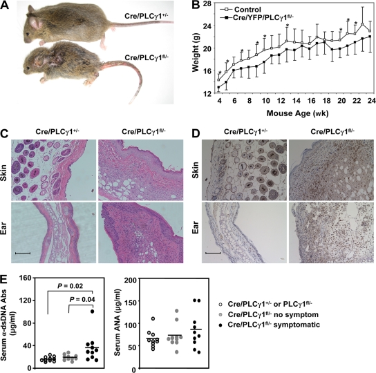
Figure 4.
Cre/PLCγ1fl/− mice develop inflammatory/autoimmune disease. (A) Cre/PLCγ1fl/− mice were smaller than littermate control mice. A pair of 6-wk-old littermates is shown. (B) PLCγ1 deficiency reduced weight gain in Cre/PLCγ1fl/− mice. Each point represents the mean weight of 7 to 14 female mice. *: P < 0.05. (C) Infiltration of inflammatory cells in the skin and ear (H&E, x200) of Cre/PLCγ1fl/− mice. Bar, 100 µm. (D) Infiltration of CD3+ T cells in the skin and ear (x200) of Cre/PLCγ1fl/− mice. The slides were analyzed by immunohistochemistry and examined by Nikon Eclipse E600. Bar, 100 µm. (E) Levels of α-double-stranded DNA antibodies and antinuclear antibodies in the serum of symptomatic Cre/PLCγ1fl/− mice compared with the indicated control mice.

