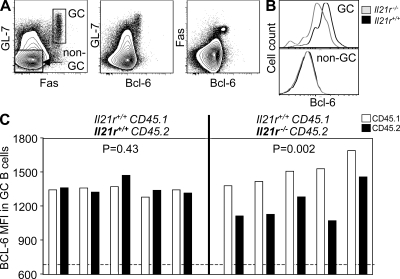
Figure 5.
Lack of IL-21R signaling reduces the expression of Bcl-6 in GC B cells. (A) Flow cytometric contour plots indicating the gating strategy for non-GC and GC B220+ cells (left) and BCL-6 expression on GL-7+ (middle) and Fas+ (right) B cells. (B and C) Histogram overlays (B) and bar graphs (C) showing the fluorescence intensity of BCL-6 staining in GC and non-GC B cells as gated in A derived from the CD45.1 Il21r+/+ or CD45.2 Il21−/− compartment of CD45.1 Il21r+/+/CD45.2 Il21r−/− mixed bone marrow chimeras 6 d after SRBC immunization. The horizontal dashed line highlights the median levels of Bcl-6 found in non-GC B cells. Each set of two bars represents the data from the CD45.1 and CD45.2 compartments of a single mouse. MFI, mean fluorescence intensity.

