
Figure 2.
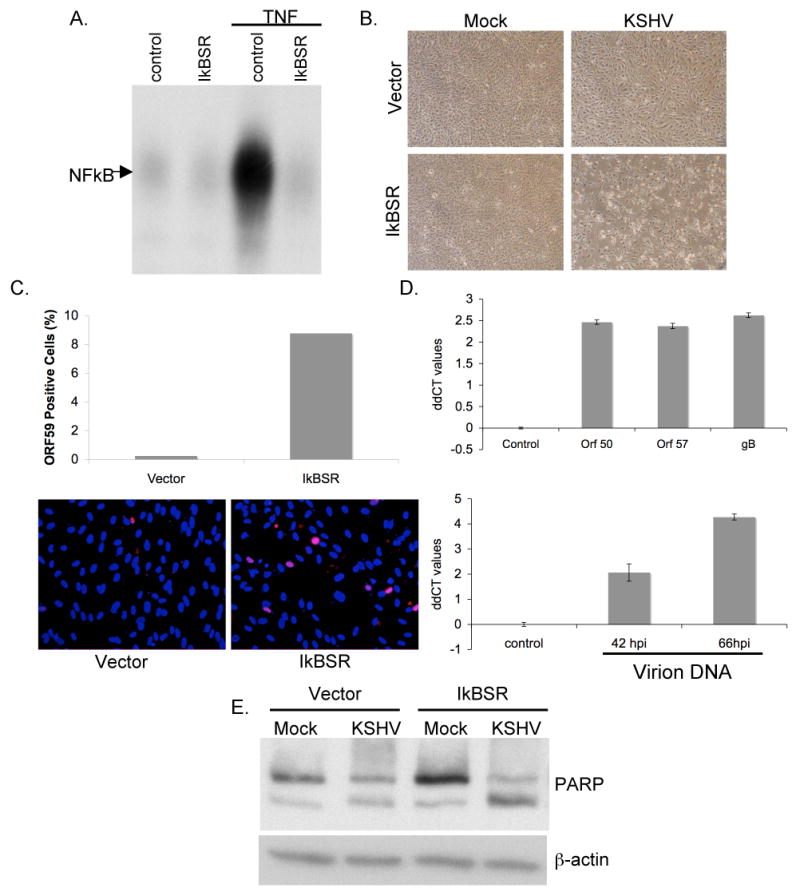
Inhibition of NFκB in HUVEC cells leads to increased cellular toxicity, increased lytic gene expression, virion production and apoptosis upon de novo infection with KSHV.
(A) HUVECs were transduced with retroviruses encoding a degradation-resistant mutant of IκBα (IκBSR). NFκB DNA binding activity in these cells was blocked upon treatment with 10ng/ml TNFα for 2hrs, as compared to those expressing vector alone.
(B) HUVECs expressing IκBSR show increased cellular toxicity upon de novo latent KSHV infection. HUVECs expressing either vector or IκBSR were infected with KSHV for 6hrs, pictures were taken at 66 hours post infection.
(C) KSHV infected IκBSR HUVECs show increased staining for ORF 59 (red) as compared to HUVECs expressing vector alone. Cells were fixed and stained at 66 hours post infection.
(D) Taqman real time PCR ddCT values (log base 2) for ORF 50, ORF 57 and gB (upper panel) and PAN promoter values (lower panel) show increased lytic gene expression and enhanced virion production in IκBSR expressing HUVECs. All values were normalized to GAPDH or spike-in DNA levels, and are expressed for each gene in IκBSR HUVECs relative to vector HUVECs. (Please see Materials and Methods sections for details on virion DNA isolation)
(E) IκBSR HUVECs show increased apoptosis upon infection with KSHV. Total protein was isolated from all cells (floating and adherent to the dish) from vector and IκBSR HUVECs at 66 hours post infection. 20ug of total protein was blotted for PARP protein and β actin as a loading control.