
Figure 4.
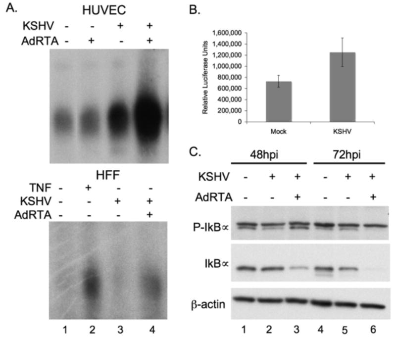
NFκB signaling is activated in lytically infected cells.
(A) Increased NFκB DNA binding activity is observed in lytically infected HUVECs (upper panel) and HFFs (lower panel), as compared to both uninfected, and latently infected cells. Nuclear fractions were harvested at 48 hours post infection, 5ug of protein was used in each binding reaction.
(B) Increased NFκB transcription is shown in 293T cells cotransfected with RTA and NFκB-luciferase reporter at 48 hours post infection. Both Mock and KSHV cells were transfected with CD8, RTA and luciferase plasmids for 36 hrs before infection, and enriched for CD8, and therefore RTA positive cells, using anti-CD8 coupled beads over magnetic columns.
(C) Lytically infected HUVECs show increased phosphorylation of IκBα (top blot, lower band) and corresponding decrease in levels of total IκBα (middle blot) demonstrating active NFkB signal transduction.