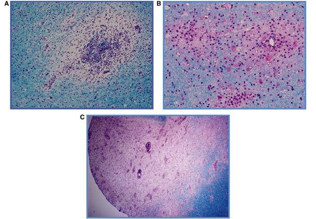
Figure 2.
Representative brain biopsies illustrating the observed patterns of demyelination. (A) Perivenous sleeve of inflammation and demyelination (20×); (B) three coalescing perivenous lesions (60×); and (C) extensive region of confluent demyelination with areas of perivenous demyelination in the periplaque white matter (4×). Luxol-fast blue periodic acid-Schiff myelin stain (A–C).

