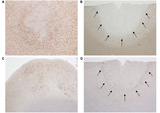
Figure 3.
Patterns of cortical pathology in perivenous demyelination cohort. (A) Perivenous intracortical demyelinated lesion (20×); (B) subpial demyelination (arrows) (4×); (C) multifocal aggregates of cortical migroglial activation (4×); (D) macrophage/microglial activation concentrated in cortical layer 3 (arrows) (4×). Immunocytochemistry for proteolipid protein (A/B) and KiM1P (C/D).

