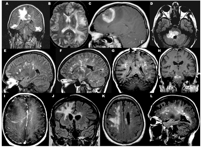
Figure 4.
Magnetic resonance imaging correlates of perivenous demyelination. (A) Coronal fluid attenuated inversion recovery (FLAIR): multifocal large ill-defined supratentorial and brainstem lesions without enhancement; (B) axial T2: large ill-defined non-enhancing uni-hemispheric lesion; (C) T1 with gadolinium: single large open ring enhancing mass with surrounding oedema; (D) axial FLAIR: large brainstem lesion with (H) punctate central and peripheral rim of enhancement; (E and F) sagittal FLAIR: numerous multifocal bilateral non-enhancing T2 lesions in subcortical white matter, basal ganglion, cerebellum; (G) coronal T1 with gadolinium: numerous bilateral enhancing subcortical white matter lesions; (I) T1 with gadolinium: faint rim of enhancement of large ill-defined lesion with mass effect; (J) coronal FLAIR; residual confluent signal change crossing the corpus callosum; (K) axial FLAIR: residual signal change oriented perpendicular to corpus callosum becoming confluent; (L) new non-enhancing periventricular white matter lesions in patient with both perivenous demyelination and confluent demyelination.

