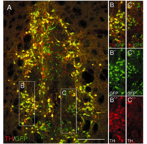
Figure 5.
Expression of the GFP reporter in intrastriatal ventral mesencephalon grafts. Immunohistochemistry for GFP (green; A–C) and TH (red; A–C) in a coronal section through the striatum from a representative animal 12 weeks after transplantation of ventral mesencephalon cells from the Pitx3WT/GFP donor group. The boxed areas are shown in greater detail on the left as individual and merged colour channels and illustrate the overlap between TH and GFP that is the case for the vast majority of GFP+ cells (B) and also the population of small TH−/GFP+ cells that tended to cluster in more central parts of the grafts (C). Scale: 200 µm.

