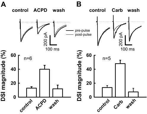
Fig. 4.
(1S,3R)-1-aminocyclopentane-1,3-dicarboxylic acid (1S,3R)-ACPD and carbachol facilitate DSI in neonatal rats (P8-9). A: traces showing DSI are all from the same neuron (top). In the presence of ACPD (10 μM) for 4–8 min, depolarization produced a robust suppression (40 ± 6% reduction of baseline eIPSC amplitude; n = 6; bottom). Prepulse trace is the average of the 5 eIPSCs just prior to a 4-s depolarization, and post pulse trace is the average of 3 eIPSCs after depolarization. B: carbachol (carb) enhances DSI in neonatal rat. Similar arrangement as in A, in the presence of carb (2 μM) for 4–8 min, the same depolarizing step leads to a 48 ± 5% reduction relative to baseline eIPSC amplitude (n = 5).

