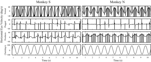
Fig. 1.
Horizontal eye velocity plotted against time during yaw-axis sinusoidal rotation at 0.5 Hz. Traces show the eye movement responses in the normal monkeys, following bilateral plugging of the lateral canals and after the prosthesis was first activated in the low-sensitivity state. Peak velocity of head rotation was 80°/s. Vestibuloocular reflex (VOR) responses were symmetric for monkey S in the normal state, but monkey N had a spontaneous nystagmus in the dark in the normal state, which measured 4.0°/s during this test session, and this biased the VOR in the positive direction. The VOR response in monkey N was symmetric, however, about this biased zero. After the prosthesis was activated, monkey N had a leftward (positive) VOR bias that reflected the small, residual spontaneous nystagmus produced by the tonic electrical stimulation of the right ear that remained after 30 min of tonic stimulation in the dark.

