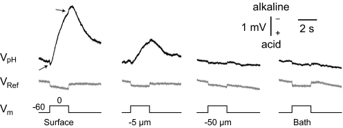
Fig. 1.
Distance profile of traces from pH-sensitive vs. saline-filled microelectrodes. Top: surface pH electrode responses to a depolarizing voltage step from −60 to 0 mV, at various distances from the cell body of a pyramidal neuron. Arrows indicate transient voltage artifacts. Middle: shows the respective profile obtained when a 2 M NaCl-filled reference pipette replaced the pH microelectrode. Note that the small, downward, rectangular deflection was common to both electrodes and present at all distances. Alkaline responses are indicated by upward deflections in this and all subsequent figures. All solutions contained benzolamide unless noted otherwise.

