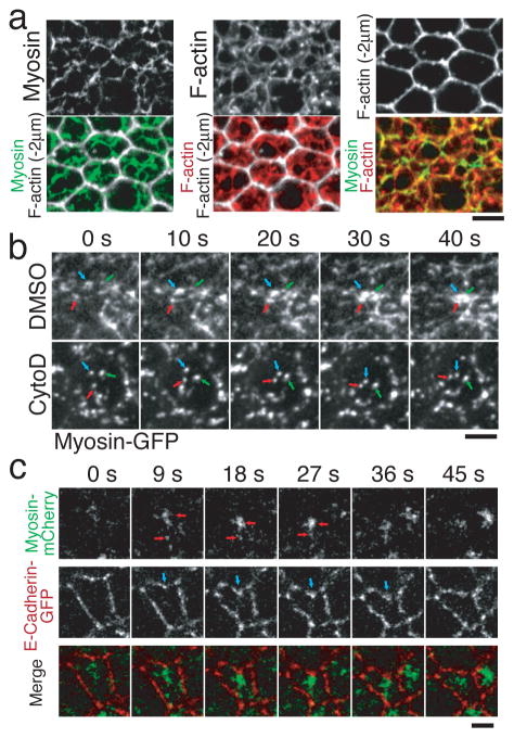
Figure 3. Pulsed myosin coalescence and adherens junction bending require an actin-myosin network.
a, Cortical myosin (green), cortical F-actin (red), and F-actin 2μm below the apical cortex (white, to illustrate cell shape) were visualized in fixed embryos. b, Timelapse images of Myosin-GFP in control injected (DMSO) and cytochalasin D (CytoD) injected embryos. Arrows indicate individual myosin spots. Note that myosin spots move, but do not coalesce in CytoD treated embryos. c, Single channel and merged timelapse images of Myosin-mCherry (green) and E-Cadherin-GFP (red). Red arrows indicate myosin coalescence. Blue arrows indicate the site where adherens junctions bend inward beneath a myosin spot. Scale bars = 4 μm.

