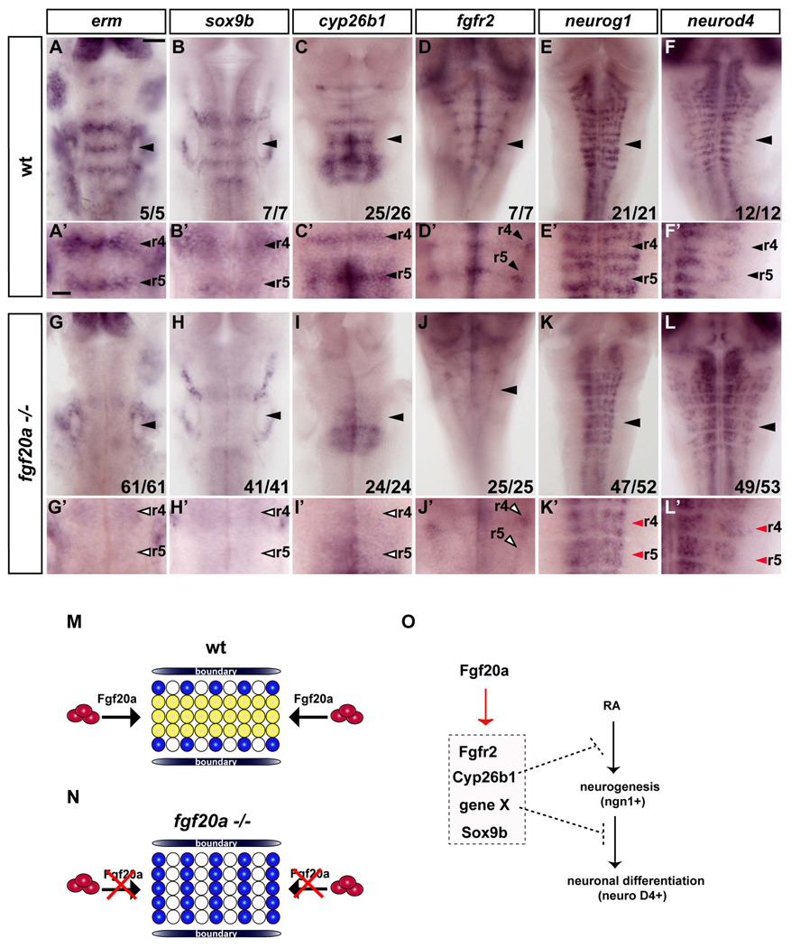
Figure 7. Fgf20a is required for inhibition of neurogenesis in segment centres.
In situ hybridisations of wild type (A–F) or fgf20a homozygous embryos (G–L) raised at 25°C. (A’–L’) show higher power images of A–L. Scale bar, 50 µm for A–L; 20 µm for A’–L’. erm expression in segment centres is significantly reduced in fgf20a mutants (open arrowheads in G’). Markers of segment centres, sox9b, cyp26b1 and fgfr2 are greatly decreased in fgf20a −/− embryos (open arrowheads in H’–J’). (K–L): fgf20a mutant embryos have ectopic neurogenesis in segment centres, detected by neurog1 (K, E) and neurod4 expression (L, F). Red arrowheads indicate ectopic neurogenesis in segment centres (K’, L’). (M–N) Model of the patterning of neurogenesis by fgf20a in hindbrain segments. In wild type embryos (M), fgf20a secreted from neurons in the adjacent mantle region (red ovals) prevents neuronal differentiation (blue circles) in segment centres by maintaining a population of progenitors (yellow circles). (N) In fgf20a mutants there is ectopic neurogenesis and low level expression of segment centre markers. (O) Summary of the regulation of genes in the non-neurogenic zone of progenitors in segment centres. fgf20a upregulates a set of genes that control different aspects of maintaining an undifferentiated population.

