
Figure 1.
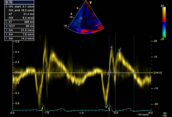
Pulsed wave tissue Doppler imaging of the RV free wall of a control subject. 1: peak myocardial systolic velocity (Sm), 2: peak early diastolic velocity (Em), 3: peak late diastolic velocity (Am) 4: isovolumetric contraction time (IVCT), 5: ejection time (ET), 6: peak myocardial isovolumetric contraction velocity (IVV), acceleration time (AT), isovolumetric acceleration (IVA).