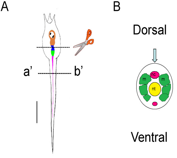
Figure 1.
Simplified diagrams showing Ciona intestinalis larval body plan and 'central nervous system'. (A) Larval body plan showing the central nervous system divisions. Orange, brain vesicle (BV) containing photoreceptive ocellus and a gravity sensing otolith (black spots). Green, presumptive motor ganglion, known variously as the visceral, trunk or tail ganglion. Pink, nerve cord (NC). The upper dotted line shown the region of the section made when preparing 'headless' larvae. The lower section on the line a'-b' shows the region of the cross section shown diagrammatically in B. (B) Diagram showing the main features of the nerve cord (pink), muscle (green) at the cross-section at the line a'-b'. The blue arrow shows the angle of view in A. This diagram is used in the following figures to show the angle of view in the micrographs. Scale bar in A, 100 μm.

