Figure 1.
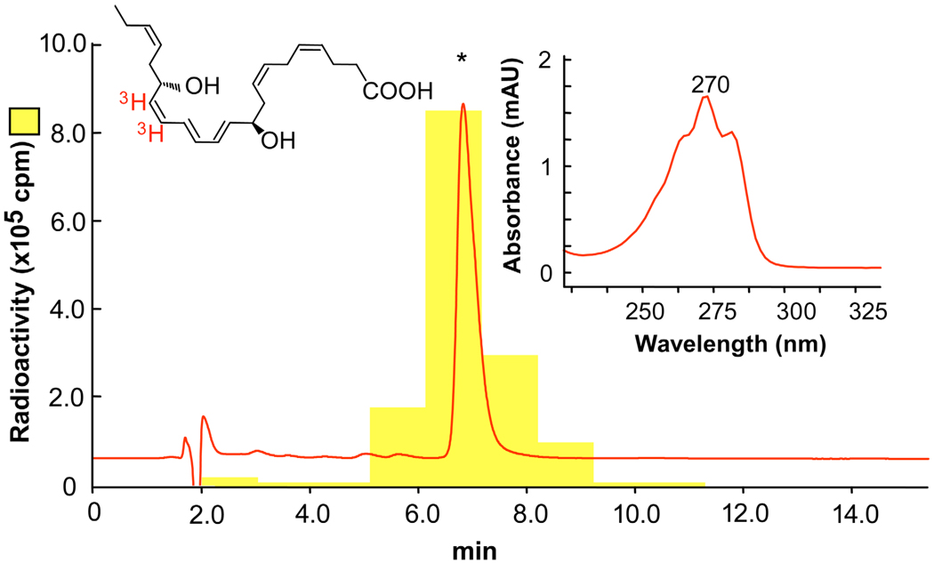
[3H]-NPD1: elution profile and UV spectrum. Reverse phase HPLC of [3H]-NPD1 was performed and recorded at 270 nm absorbance, showing identical retention time (asterisk) as standard NPD1. The locations of the [3H] in red are indicated on the chemical structure of NPD1. Insert shows the characteristic online UV spectrum.

