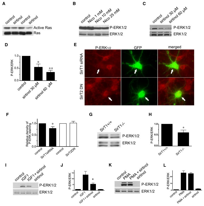
Figure 2.
Inhibition of SirT1 deacetylase decreases Ras/ERK1/2 activation in cultured neurons and in vivo. (A) Representative blots showing the effect of SirT1 inhibitor sirtinol on Ras activation. 10–14 DIV neurons were treated with vehicle or 60 μM sirtinol for 4 hrs and subjected to Ras activation assay or immunoblotted with anti-Ras. (B, C) Representative blots showing the effect of SirT1 inhibitors nicotinamide (B) and sirtinol (C) on ERK1/2 signaling. 10–14 DIV neurons were treated with Nico for 48 hrs or sirtinol for 4 hrs. Total cell lysates were collected for SDS-PAGE and blotted with anti-phospho-ERK1/2 and anti-ERK1/2, respectively. (D) Quantification of immunoblots showing the effect of sirtinol on ERK1/2 activation. (E) 7 DIV cortical neurons were transfected with U6Pro-SirT1-siRNA+GFP or dominant-negative GFP-SirT2 (SirT2DN) and 48 hrs later were immunostained with anti-phospho-ERK1/2. (F) Quantification of immunodensity from 12 transfected and 12 nearby non-transfected cells for SirT1 siRNA and SirT2DN, respectively. (G, H) Representative blots (G) and quantification (H) showing the effect of SirT1 deacetylase on ERK1/2 signaling in mice hippocampus. (I, J, K, L) Representative blots (I, K) and quantification (J, L) showing the effect of sirtinol on ERK1/2 signaling in HEK cells. HEK cells were starved for 15 hrs in media containing 0.5% FBS and incubated with 60 μM sirtinol for 4 hrs and then treated with IGF-1 or PMA for 5 min. Cell lysates were subjected to SDS-PAGE and blotted with anti-phospho-ERK1/2 or anti-ERK1/2. Quantifications are shown as mean±SEM.

