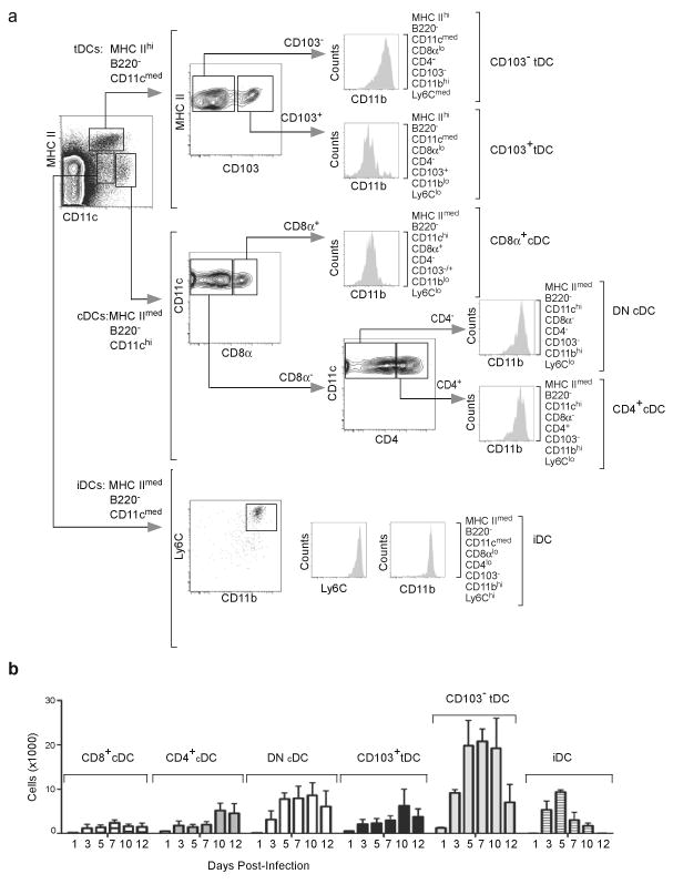
Figure 2. DC subsets in mLN after influenza infection.

(a) Mice were infected intranasally with PR8 and DC subsets were analyzed by flow cytometry on day 7 after infection. Plasmacytoid DCs were initially excluded based on B220 expression and the three major subsets that remained were defined as conventional DCs (cDCs: MHCIImedCD11chi), tissue DCs (tDCs: MHCIIhiCD11cmed) and inflammatory DCs (iDCs: MHCIImedCD11cmedLy6chi). Each of these populations was further characterized based on the expression of CD8α, CD4, CD103, CD11b and Ly6C.
(b) C57BL/6 mice were infected intranasally with PR8 and CD8α+ cDCs, CD4+CD8α−cDCs, CD8−CD4−cDCs, CD103+CD11blo tDCs, CD103−CD11bhi tDCs and iDCs were enumerated by flow cytometry as described in a. Values represent the total numbers (mean ± SD) of each subset calculated at each time point (n = 4–5 mice / time point). Data are representative of three independent experiments.
