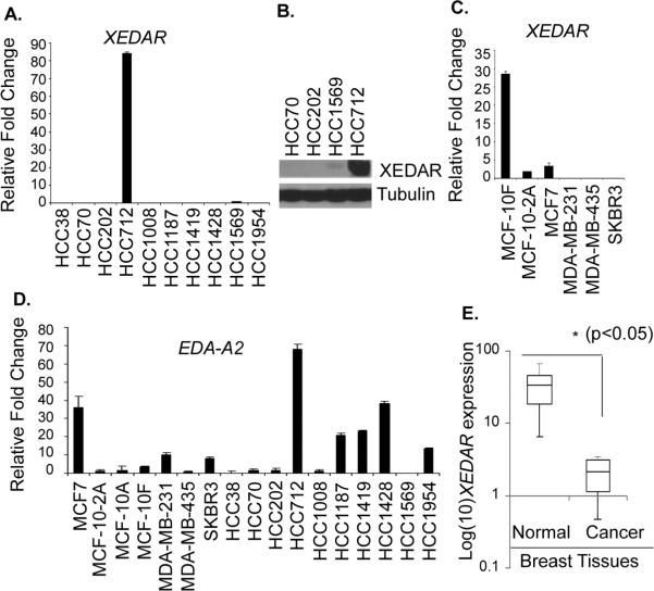
Figure 1. Down regulation of human XEDAR expression in breast cancer cell lines and tissues.
A. Expression of XEDAR was determined by qRT-PCR and normalized to β-actin as house keeping control in 10 human breast cancer cell lines obtained from Dr. A. F. Gazdar. PCR reactions were performed in triplicate and the data presented as fold change in target gene expression (Mean ± S.E.) over the negative control sample, as explained in the Material and Methods section. XEDAR expression was observed in either non-tumorigenic cell lines derived from mammary epithelial cells or in normal breast samples, but not in tumorigenic or highly metastatic breast cancer cell lines. B. Western blot showing high level expression of XEDAR protein in the HCC712 cells. Tubulin was used as an internal loading control. C. XEDAR mRNA expression, as determined by qRT-PCR, in an additional panel of 6 breast cancer cell lines representing different stages of differentiation. D. qRT-PCR analysis of EDA-A2 (XEDAR ligand) showing its more ubiquitous expression than XEDAR in various breast cancer cell lines. E. Box-Whisker plot of XEDAR expression in breast cancer tumors (n=10) and corresponding benign tissues (n=6). The level of XEDAR expression was significantly higher in benign tissues as compared to tumor samples (*P<0.05; Mann-Whitney U test).

