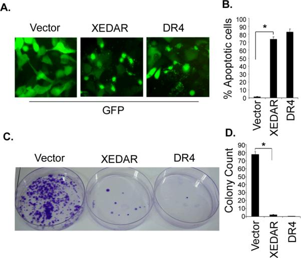
Figure 5. XEDAR induces cell death.
A–B. MDA-MB-231 cells were transfected with indicated plasmids (1 μg) along with a green fluorescent protein-encoding plasmid (GFP, 200 ng) using Lipofectamine as described in Material and Methods. Cells were examined under a fluorescence microscope and photographed 48 h after transfection. XEDAR- and DR4-transfected cells had a rounded appearance and were detaching from the plate, whereas the cells transfected with the vector have a normal morphology. C–D. MDA-MB-231 cells were transfected with the indicated plasmids as described in Figure 5A. The cells were plated on fresh medium and selected with Zeocin (200 μg/ml). After 14 days of drug selection, the cells were fixed and stained with crystal violet and the numbers of colonies were counted.

