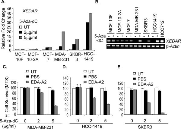
Figure 6. 5-Aza-dC reinduces XEDAR expression and sensitizes breast cancer cell lines to EDA-A2 induced cell death.
A. The indicated human breast cancer cell lines were treated with two different concentrations (2 and 5 μg/ml) of methyltransferase inhibitor 5-Aza-deoxycytidine (5-Aza-dC) for 4 days and restoration of XEDAR expression was analyzed by qRT-PCR and shown as fold increase over the untreated control cells. Re-expression of XEDAR was observed in the cell lines MDA-MB-231, SKBR3, and HCC1419 with methylated XEDAR promoter in the basal state. B. Representative qRT-PCR data of cell lines treated with 5μg/ml 5-Aza-dC showing re-expression of XEDAR. β-actin was using as internal loading control for equal amount of cDNA in PCR reaction. cDNA from HCC712 was used as positive control for XEDAR amplification. C–E. The indicated cell lines with methylated XEDAR promoter were treated for 4 days with 2 and 5μg/ml of 5-Aza-dC. The cells were then treated with recombinant EDA-A2 (100 ng/ml) for 72 h and cell survival determined by MTS assay as described previously (37). The untreated cells served as a negative control and PBS was used as an EDA-A2 vehicle control. Increasing dose of 5-Aza-dC treatment caused decreased cell survival upon treatment with EDA-A2.

