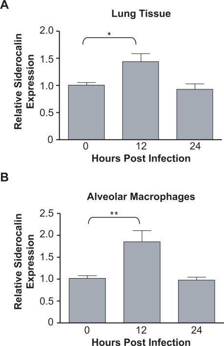
FIGURE 2. Siderocalin is induced in lung tissue and alveolar macrophages following M.tb infection.
Seven to nine week old female C57BL/6J mice were infected with the Erdmann strain of M.tb via aerosol. (A) Lungs were surgically removed 12 and 24 h post-infection and homogenized in TRIZOL to prepare total RNA. (B) Bronchoalveolar lavage fluid was collected at 12 and 24 h after infection and alveolar macrophages prepared. The cells were homogenized in TRIZOL. Siderocalin expression was determined in total lung and alveolar macrophages using qRT-PCR. Gene expression corresponds to the 2−ΔΔCT between siderocalin and a reference gene (36B4). For statistical analysis, the means ± SEM from a single experiment (n=5) for lung and alveolar macrophages are shown. (*) denotes P ≤ 0.05, (**) denotes P ≤ 0.01 using the Student's t test.

