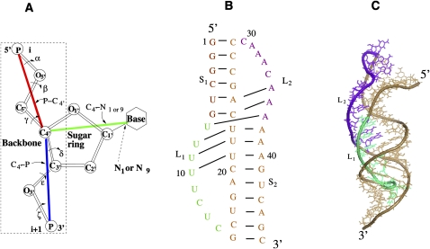
FIGURE 1.
(A) Virtual bonds for a nucleotide: 5′P–C4′3′, 5′C4′–P3′, and C4′–N1 (primidine) or C4′–N9 (purine). (B) The secondary structure for hTR pseudoknot (Theimer et al. 2005). (C) The three-dimensional structure of the hTR pseudoknot (PDB code: 1YMO) (Theimer et al. 2005). Loops L1 and L2 span across the major and minor grooves of helices S2 and S1, respectively.

