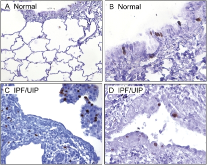
Figure 7.
Ki-67 staining in serial sections of lung tissues in IPF/UIP. (A) Low magnification of Ki-67 staining indicating sparse staining of alveolar epithelial cells and columnar epithelial cells of airway walls (×10 objective). (B) High magnification image a section of a small airway showing Ki-67+ columnar epithelial cells (×40 objective). (C and D) High magnification images of Ki-67+ alveolar epithelial cells in lung tissues from patients with IPF/UIP. Note the paucity of Ki-67 staining in the fibroblastic focus (centrally located) in C and the abundant staining in a region of alveolar epithelial cell hyperplasia (D). C: ×20 objective; D: ×40 objective.

