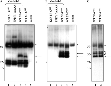
Figure 5.
Ubiquitination of proSP-C. HEK293 cells were cotransfected with plasmids encoding WT (SP-C1–197) or mutant (K6R and PPDY/AAAA) proSP-C and plasmid encoding full-length human Nedd4-2 (+Nedd4-2). Cell lysates were prepared 24 hours after transfection and immunoprecipitated for proSP-C. Antigen–antibody complexes were captured by protein G sepharose and analyzed by SDS-PAGE/Western blotting with antibody directed against the N-terminal peptide of proSP-C (A), or antibody that detects mono- and polyubiquitinated proteins (B). Overexposure was required to detect ubiquitinated SP-C in (A) and (B) was deliberately overexposed to demonstrate absence of polyubiquitination. (C) The experiment was repeated, and lysates from cells transfected with WT SP-C were analyzed as in (B). A shorter exposure confirmed the presence of two major ubiquitinated forms of SP-C (long arrows) and two minor forms (arrowheads). Short arrows indicate proSP-C; *IgG detected by secondary antibody.

