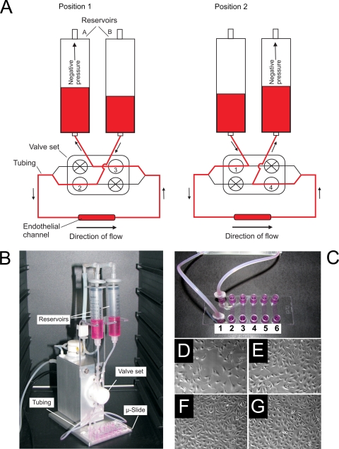
Fig. 1.
The circulatory model. (A) Schematic representation of the circulatory model. Air pressure in the reservoirs and opening/closing of the valve set are controlled via an air pump and computer software. Note that in both position 1 and position 2 unidirectional flow through an endothelial channel is maintained. (B) Picture of the circulatory model (ibidi pump system) with reservoirs, valve set, tubing, and μ-Slide labeled. (C) Closeup picture of the μ-Slide. Note that each μ-Slide houses six independent channels, which can be seeded with endothelial cells and connected to the circulatory model via Luer adaptors. (D to G) Micrograph images of the development of endothelial cells within a μ-Slide channel, 1 (D), 2 (E), and 3 (F) days postseeding and following 20 min under flow (G). Note that monolayers are confluent by 3 days postincubation and that 20 min flow does not overtly effect the morphology of the endothelial monolayer.

