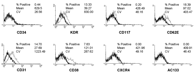
Fig. 2.
Flow cytometric analysis of endothelial cells obtained from CD34+ cells after 4 weeks of culture. The dark line identifies the cells labeled with isotype control antibody and the faint line identifies the cells labeled with antibodies specific for the surface markers. Cultured cells were positive for KDR, CD31 and CD62E, but negative for CD34, CD117, AC133 and CXCR4 (cut off, 10%).

