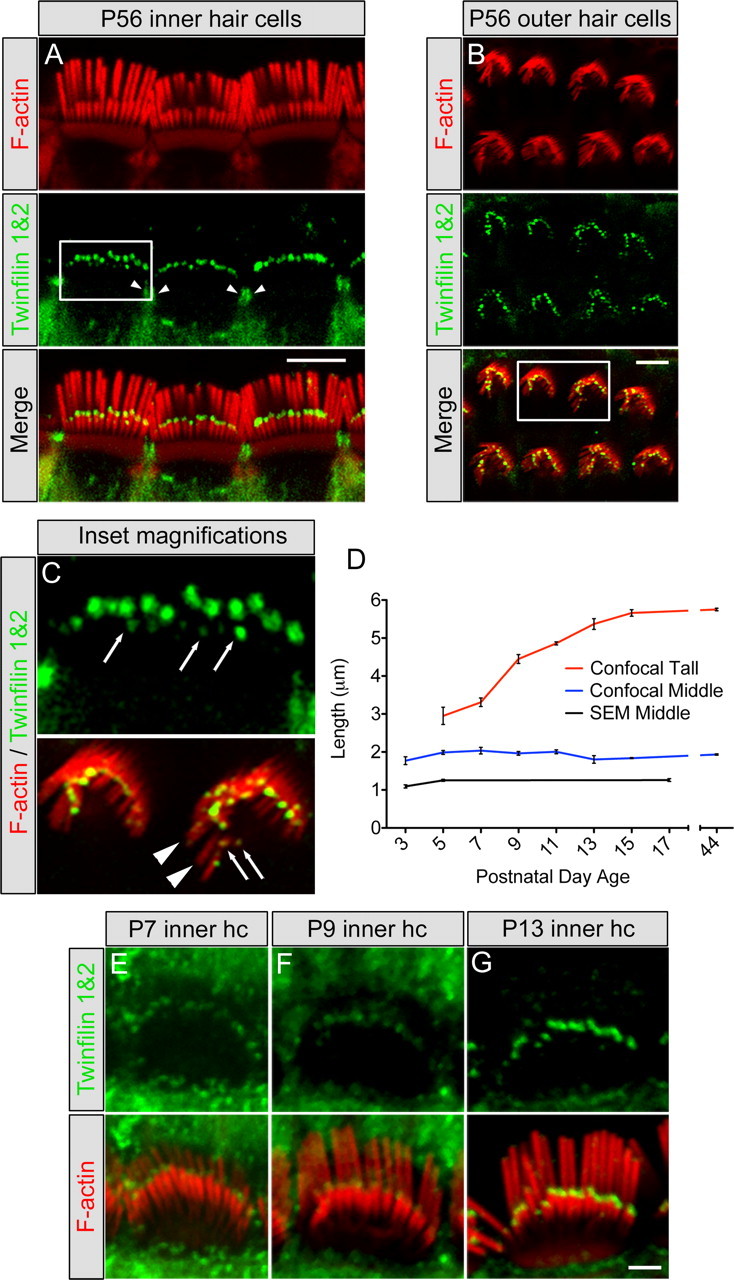
Figure 2.

Twinfilin 2 localization in hair cell stereocilia and developmental characterization. A, B, Localization using a twinfilin 1&2 antibody (green) on P56 mouse cochlea showing tip staining of the middle and short rows of stereocilia in inner hair cells (A) and outer hair cells (B) as well as the pericuticular necklace region of inner hair cells (arrowheads). F-actin was labeled with TRITC-conjugated phalloidin (red). C, Top, Enlargement of the square in A showing the staining at the tips of short-row stereocilia in inner hair cells (arrows); bottom, enlargement of the square in B showing lack of staining at tips of tall-row stereocilia (arrowheads) and staining at the tips of short-row stereocilia (arrows). D, Inner hair cell stereocilia lengths for tall (red line) and middle (blue line) rows using confocal microscopy, and middle-row lengths (black line) determined with scanning electron microscopy (SEM) (mean ± SD, n = 3). Scanning electron microscopy-measured lengths were consistently shorter than confocal-measured lengths, likely due to the dehydration steps in tissue processing. E–G, Developmental expression of twinfilin 2 (green) shows increasing labeling intensity with age from P7 (E) to P9 (F) to P13 (G). Scale bars: = A, B; 5 μm; E–G, 2 μm.
