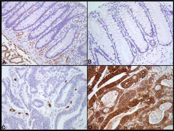
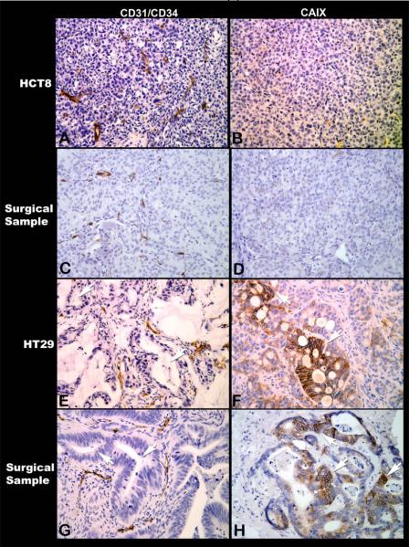
Figure 1.
Microphotographs of CD31/CD34 immunostaining to visualize microvessels (left panels) and CAIX immunostaining to demonstrate tumor hypoxic regions (right panels) (All magnifications, X200). [1(i)] Normal colon mucosa (A) with microvessels (brown) uniformly distributed except for the glandular structures that do not contain vessels and is yet not hypoxic (B). In contrast, adenocarcinoma do not contain microvessels in the glandular regions, but only in the surrounding stroma (C) and the cancer cells are strongly positive for hypoxia marker CAIX (brown) in many such glandular regions (D). [1(ii)] Photomicrographs of HCT-8 and HT-29 human colon cancer xenografts along with human surgical samples of colorectal cancers with similar histomorphological characteristics. Poorly differentiated HCT-8 is uniformly well vascularised (A) and has no regions of hypoxia (B). Similar vascular arrangement (C) and absence of hypoxia (D) are seen in this surgical sample of poorly differentiated colorectal cancer. In contrast, in HT-29, the cancer cells form glandular structures (E, arrows) that being avascular are hypoxic (F, arrows). Most surgical samples of colorectal cancers have such glandular structures (G, arrows) containing hypoxic cancer cells within the glandular regions (H, arrows).


