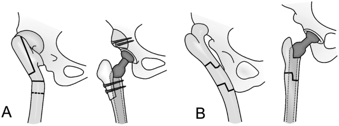
Figure 2.
Osteotomies used for various deformities of the femur. The femoral shaft is usually transected distal to the lesser trochanter, as shown by the transverse solid line in (A). A dotted line demonstrates the most distal possible level of the osteotomy. A. Proximal shortening osteotomy with distal advancement of the greater trochanter (vertical solid line) in hips with a previous proximal Schanz osteotomy. B. Segmental shortening with angular correction for hips with a previous, more distal Schanz osteotomy. Copyright for the illustrations in this figure is owned by The Journal of Bone and Joint Surgery, Inc. (published in Eskelinen et al. Cementless total hip arthroplasty in patients with high congenital hip dislocation, J Bone Joint Surg Am. 2006; 88: 80-91). Reproduced with permission.

