
Figure 2.
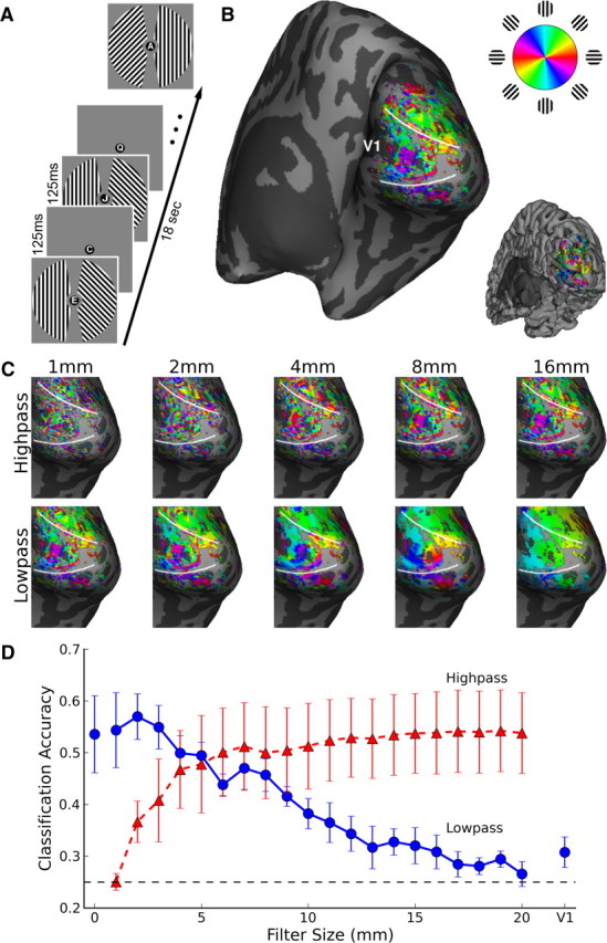
Orientation information at multiple spatial scales in human visual cortex. A, Human subjects viewed 18 s blocks of independent oriented gratings in the right and left visual fields. B, Unthresholded orientation preference of individual voxels rendered on the inflated cortical surface of the right hemisphere of a representative subject, with all visually responsive (p < 0.01) voxels shown. Inset shows the folded cortical surface from the same viewpoint. C, Fine- and coarse-scale activity patterns revealed by ideal volumetric highpass and lowpass filtering. D, Orientation classification accuracy plotted as a function of filter size for highpassed (red triangles) and lowpassed (blue circles) data. Leftmost blue circle, classification of original data; rightmost blue circle, classification of mean activity in contralateral V1. Error bars indicate ±1 SEM based on N = 4 subjects. Dashed line indicates chance-level (25%) performance.