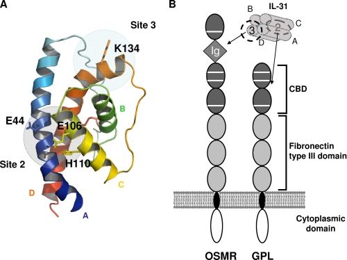
FIGURE 7.
Model representation of IL-31. A, selected model of human IL-31 predicted by I-TASSER. α A, B, C, and D helices are colored in blue, green, yellow, and orange, respectively. The predicted disulfide bridge is highlighted in red. Binding sites 2 and 3 are indicated by gray circles. B, schematic view of the domain structure of IL-31, OSMR, and GPL and representation of the IL-31/IL-31 receptor complex interaction. Gray square, Ig-like domain; dark gray oval, CBD; light gray oval, fibronectin type III domain.

