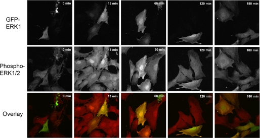
FIGURE 1.
Localization of GFP-ERK1 in MEFERK1−/− cells. MEFERK1−/− cells expressing GFP-ERK1 (top row, green in overlay) and endogenous ERK2 were fixed in methanol at −20 °C after starvation for 12 h (time 0) and at different time points after activation by addition of serum. Cells were stained for phosphorylated ERK1/2 (middle row, red in overlay).

