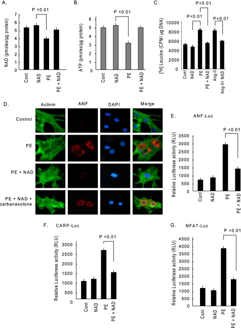
FIGURE 2.
NAD treatment blocks cardiac hypertrophic response in vitro. A, cardiomyocytes were treated with PE either alone or together with NAD. After 48 h of treatment cells were harvested and NAD content measured. B, ATP content was measured in the same groups of cells. C, cardiomyocytes were treated with PE (20 μm) or Ang-II (2 μm) in the presence or absence of NAD and labeled with [3H]leucine. Forty-eight hours after treatment, cells were harvested, and [3H]leucine incorporation into total cellular protein was measured. D, cardiomyocytes were stimulated with PE and then treated with NAD, either alone or together with carbenoxolone (10 μm). Cardiomyocytes were identified by α-actinin staining using anti-α-actinin antibody (green). The release of ANF from nuclei was determined by staining cells with an anti-ANF antibody (red). 4′,6-Diamidino-2-phenylindole (DAPI) stain was used to mark the position of nuclei. E and F, cardiomyocytes were transfected with luciferase reporter plasmids responsive to ANF (E) or CARP (F). Luciferase activity was measured after 8 h of treatment with PE, in the presence or absence of NAD (G). Cardiomyocytes were infected with a NFAT responsive luciferase reporter adenovirus vector. Twelve hours after infection cells were treated with PE in the presence or absence of NAD, and luciferase activity was determined 8 h post-treatment. Cont., control. Values presented in histograms are mean ± S.E. of six experiments.

