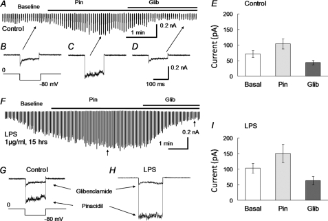
FIGURE 2.
Augmentation of KATP currents with LPS incubation. Whole cell voltage clamp was performed in freshly dissociated aortic SMCs. The bath solutions contained 145 mm K+. The same solution was used in the recording pipette with addition of 1 mm ATP, 0.5 mm ADP, and 1 mm free Mg2+. A, in a control experiment, small inward currents were seen upon the formation of the whole cell configuration. The currents were increased by pinacidil (10 μm). The maximal activation was reached in 2 min, whereas glibenclamide (10 μm) reduced currents to a level even below the baseline. B–D, individual records of inward currents at baseline (B) and with pinacidil (Pin, C) or glibenclamide (Glib, D). E, summary of the current amplitude under these conditions (n = 21 cells). F, the pinacidil- and glibenclamide-sensitive currents were studied in another SMC that had been treated with LPS (1 μg/ml) overnight. The current amplitude increased significantly after the whole cell patch formation, presumably produced by intracellular dialysis of ADP and Mg2+. The currents were further augmented by pinacidil, reaching a peak that doubled that without LPS treatment in A. The pinacidil-activated currents were completely suppressed by glibenclamide. G, superimposed are individual currents obtained from C and D. H, current traces were similarly taken from F at the position indicated by arrows. I, effect of LPS on whole cell currents of dissociated SMCs (n = 17 cells).

