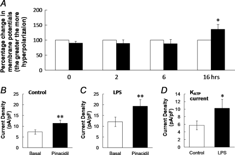
FIGURE 3.
A, effect of LPS on membrane potentials (Vm) was studied in vascular SMCs freshly dissociated from the mouse aorta by parallel comparison of the Vm recorded from cells treated with and without LPS. Although no obvious changes in Vm were seen with an LPS treatment for 0, 2, and 6 h, significant hyperpolarization occurred with a 16 h exposure (*, p < 0.05; n = 10). B–D, enhancement of current density with LPS exposure. The current density was calculated by dividing the current amplitude by the whole cell capacitance in each cell. Without LPS, the pinacidil-activated current (I) density was ∼54% greater than the basal currents (B, n = 21, **, p < 0.01). After an overnight treatment with LPS (1 μg/ml), the density of basal currents increased by 50%, whereas the pinacidil-activated current density was further elevated by 59% (C, n = 17). KATP currents were isolated by subtraction of the glibenclamide-sensitive currents from the pinacidil-activated currents (Fig. 2, G and H). The KATP current density was significantly enhanced after LPS exposure (*, p < 0.05, D).

